
Jessie Thomson
Osteoarthritis is a degenerative disease that effects the entire joint, degrading articular cartilage and deforming the surrounding bones and tissue of the affected joint. The disease affected 8.5 million people in the UK in 2012, and caused over £2.6 billion annually in analysing and repairing the joint, and alleviating the pain caused. Osteoarthritis is a severely debilitating disease across the population. With no known pathogenesis, research emphasis lies with finding new and improved ways of supplementing the effects of the disease.
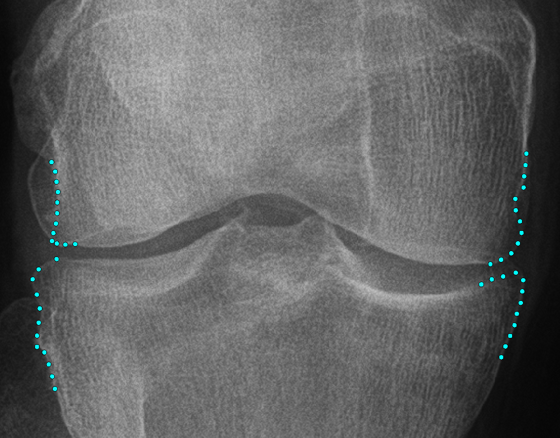
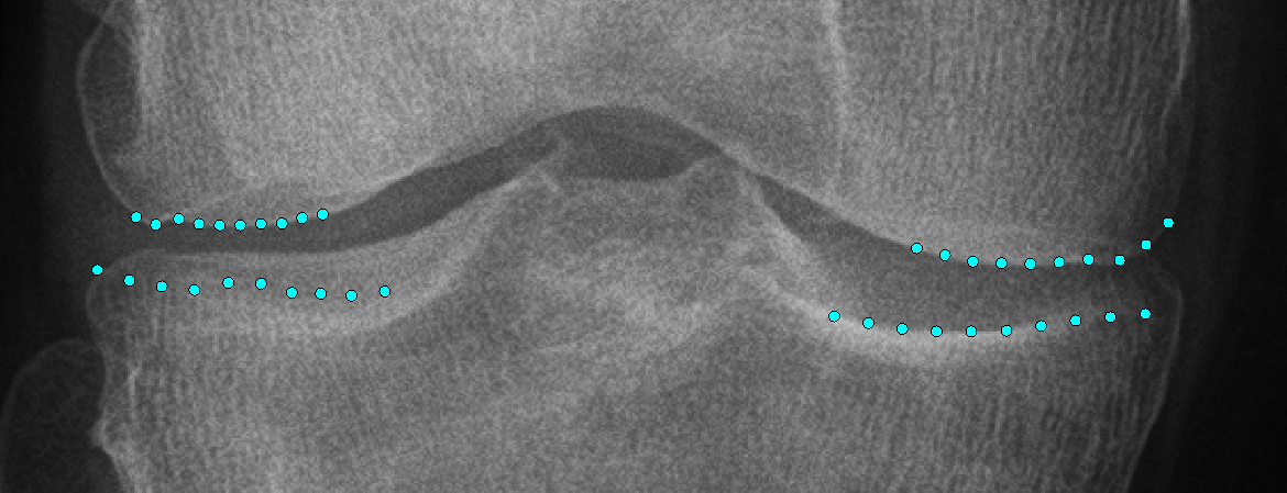
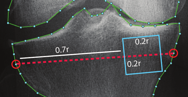
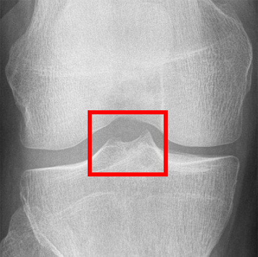
This study looks at the area of the knee, which is one of the main areas afflicted by Osteoarthritis. The project aims to analyse the mechanics of the disease in the joint to identify significant markers for Osteoarthritis and related knee pain. These markers will be based on various shape and texture features extracted across the whole of the joint. Current clinical methods rely on manually applied, semi-quantitative descriptions of the osteoarthritic features to detail the severity of the disease. Due to the semi-quantitative nature of these methods and the weighting of multiple OA features per grade, this can often be at risk of subjective beliefs of the grader. Leading to discerpencies and reliability issues when clinicans are analysing large sets of radiographics images. The application of automated methods helps to solve this issue with subjectivity, through the use of standardised, quantitative, rules and measurements. Due to the multifactorial nature of Osteoarthritis, the project looks at multiple explicit and implicit features relater to OA. These features include: overall shape, osteophytes, trabecular structure, tibial spines and inter-condylar notch, and joint space.
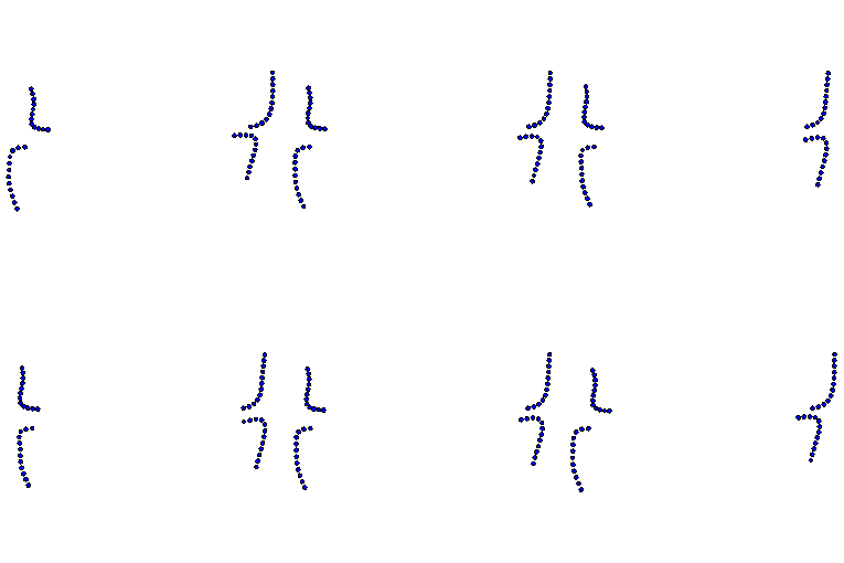
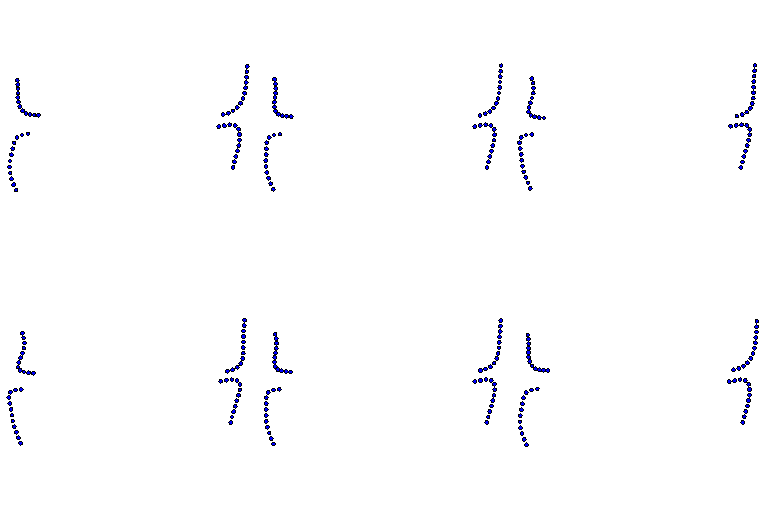
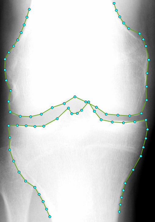
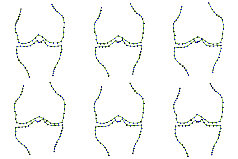
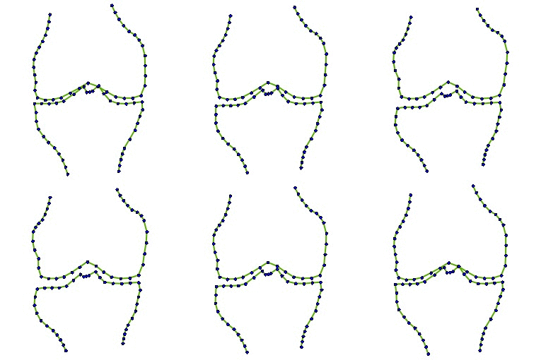
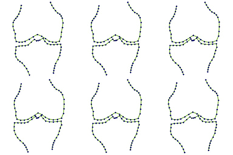
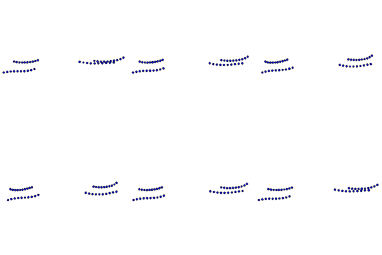
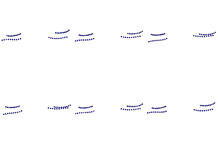
The project uses a 74 point model manually annotated on 500 images (37 points each for both the Tibia and Femur) and an RFCLM algorithm to find these points in a new image.




| Experiment | AUC (95% CI) |
|---|---|
| Current OA | 0.9035 (0.897 - 0.91) |
| Current pain | 0.6629 (0.65 - 0.675) |
| Later onset OA | 0.6137 (0.587 - 0.64) |
| Later onset pain | 0.6085 (0.594 - 0.623) |
Automated shape and texture analysis for detection of Osteoarthritis from radiographs of the knee, Proc. MICCAI 2015, Part 2, pp.127-134
J. Thomson, M. Parkes, D. Felson, T. ONeill and T. F. Cootes, Automated multi-feature analysis of current and future onset pain in osteoarthritic knees
,
Int. Workshop on Osteoarthritis Imaging, (abstract).
J. Thomson, T. ONeill, D. Felson and T. F. Cootes, Detecting osteophytes in radiographs of the knee to diagnose Osteoarthritis
Proc. MICCAI MLMI
2016 (to appear).
Under review: L. Minciullo, J. Thomson, T. F. Cootes, Combination of Lateral and PA View radiographs to Study Development of Knee OA and Associated Pain
,
SPIE 2016
In preparation: J. Thomson, M. Parkes, D. Felson, T. ONeill and T. F. Cootes, Automated Radiographic Multi-feature Analysis of Current and Future Onset OA
and Pain
, Osteoarthritis and Cartilage