Integrated Brain Image modelling (IBIM)

Research Associate: Kola Babalola

Principal Investigator: Tim Cootes

Project Period: September 2004 - October 2007

Contents

  1. Introduction
  2. Profile Active Appearance Models
  3. Application to segmentation
  4. Evaluation of different segmentation methods
  5. Associated software
  6. Selected publications
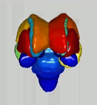
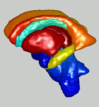
A view of the surface of some of the subcortical structures   A view of the surface of some of the subcortical structures

Introduction

This was an EPSRC funded project carried out by the Imaging Science and Biomedical Engineering (ISBE) department of the University of Manchester in collaboration with the following groups from three other major UK universities.

The overall aim of the project was to produce systems for analysing structural brain images, to aid segmentation of brain substructures and quantification of how they relate to each other in relative size, position and shape.

Each group developed their methods independently. All groups had access to a rich dataset of over 300 images with a standardised protocol for training and testing the methods. This enabled quantitative evaluation of the four different methods over a large dataset of 270 images - one of the unique characteristics of this project.

At Manchester our approach was based on profile Active Appearance Models. We also coordinated the evaluation of the different methods.

Back to contents

3D Profile Active Appearance Models

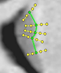
We used a version of Active Appearance Models (AAMs) that models texture sampled at profiles normal to a surface.

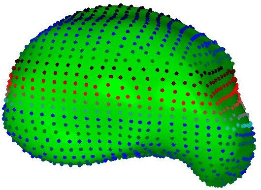
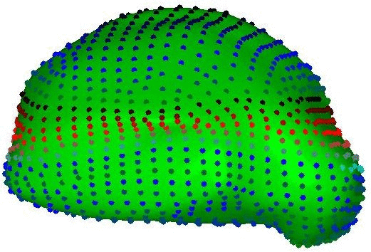
The construction process involved the identification of homologous points across the image — the so called correspondence problem. We addressed this using groupwise registration of binary images to produce surface meshes approximating the structure(s) of interest for each member of the training set. Profiles were sampled at the vertices of these meshes.

Surface mesh of the thalamus of a subject Surface mesh of the thalamus of a subject Schematic illustrating sampling of a profile across a surface
Back to contents

Application to segmentation

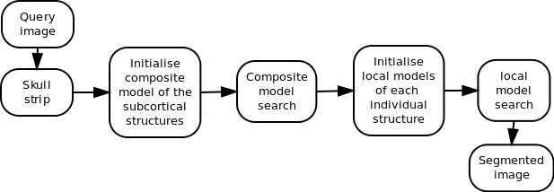
We constructed a composite profile AAM of all the structures we were interested in, and individual models of each separate structure. The composite is initialised in the image using affine registration and the initial segmentation it produces is refined by the models of the individual structures.

Schematic diagram of the steps involved in segmentation
Back to contents

Evaluation of the four segmentation methods

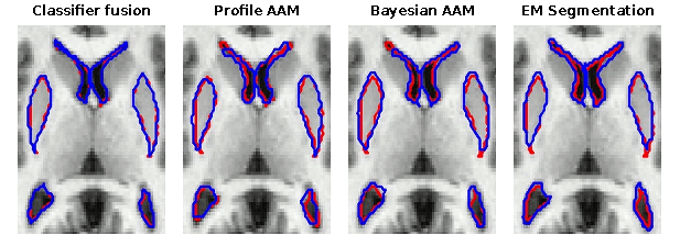
The four methods developed across the different sites were comprehensively evaluated on a dataset of 270 images. More details on this are available here.

Image comparing results obtained by AAM with those obtained by AAM after post-processing with the regressor
Back to contents

Associated Software

Executables for 3D AAMs to segment 10 subcortical structures are available here.

Back to contents

Selected publications

An evaluation of four automatic methods of segmenting the subcortical structures in the brain. KO Babalola, B Patenaude, P Aljabar, J Schnabel, D Kennedy, W Crum, S Smith, T Cootes, M Jenkinson, D Rueckert. Neuroimage, vol. 47, pp. 1435-1447, October 2009, doi:10.1016/j.neuroimage.2009.05.029

Comparison and Evaluation of Segmentation Techniques for Subcortical Structures in Brain MRI. KO Babalola, B Patenaude, P Aljabar, J Schnabel, D Kennedy, W Crum, S Smith, TF Cootes, M Jenkinson, and D Rueckert. In Proceedings of the 11th International Conference on Medical Image Computing and Computer Assisted Intervention (MICCAI), Lecture Notes in Computer Science, vol. 5241, pp. 409-416, September 2008, doi: 10.1007/978-3-540-85988-8_49

Comparing the Similarity of Statistical Shape Models using the Bhattacharya Metric. KO Babalola, TF Cootes, B Patenaude, A Rao, and M Jenkinson. In Proceedings of the 9th International Conference on Medical Image Computing and Computer Assisted Intervention (MICCAI), Lecture Notes in Computer Science, vol. 4190, pp. 142-150, October 2006, doi: 10.1007/11866565_18

Cannonical Correlation Analysis of Sub-cortical Brain Structures Using Non-rigid Registration. A Rao, Kolawole Babalola, and Daniel Rueckert. In Proceedings of the 3rd International Workshop on Biomedical Image Registration (WBIR), Lecture Notes in Computer Science, vol. 4057, pp. 66-74, July 2006, doi: 10.1007/11784012_9

Registering Richly Labelled 3D Images. KO Babalola and TF Cootes. In Proceedings of the International Symposium on Biomedical Images, Washington, Virginia, USA, April 2006, 10.1109/ISBI.2006.1625056

Back to contents