
Nailfold Capillaroscopy
NCM Capture Software
- For a demonstration of the new nailfold capillaroscopy system developed at the University of Manchester click here.
- For help and information on using the NCM software we have developed click here here.
Research overview
Nailfold capillaroscopy is technique used to capture images of capillaries in the fold of skin immediately below the fingernail (the nailfold). Here the capillaries lie parallel to the skin and can be visualised using visible light (with green light providing the best contrast between red blood cells and the surrounding the tissue). As such the nailfold capillaroscopy provides a non-invasive, cost-effective method of visualising the human microvasculature system in real-time.
It may be used in many clinical situations, but is particularly helpful in diagnosing and tracking the progression of connective diseases that affect the structure and function of capillaries.
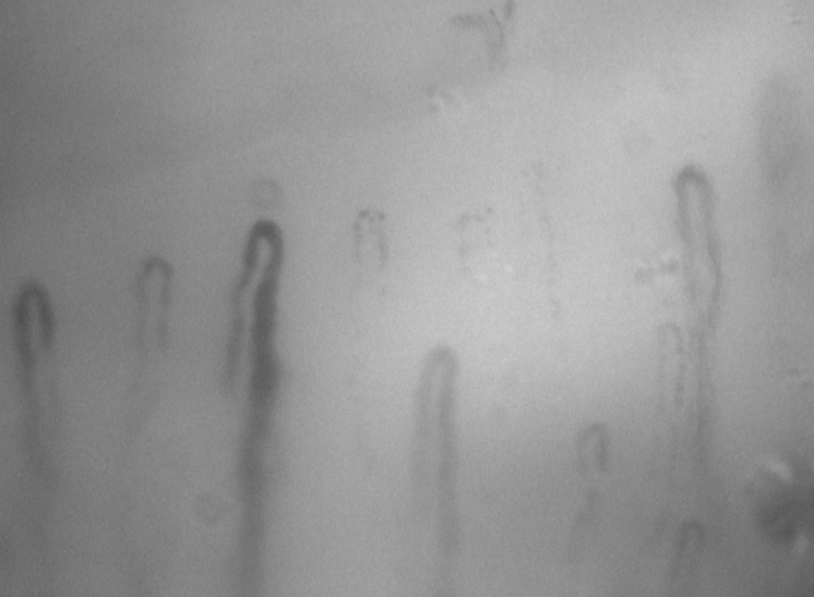
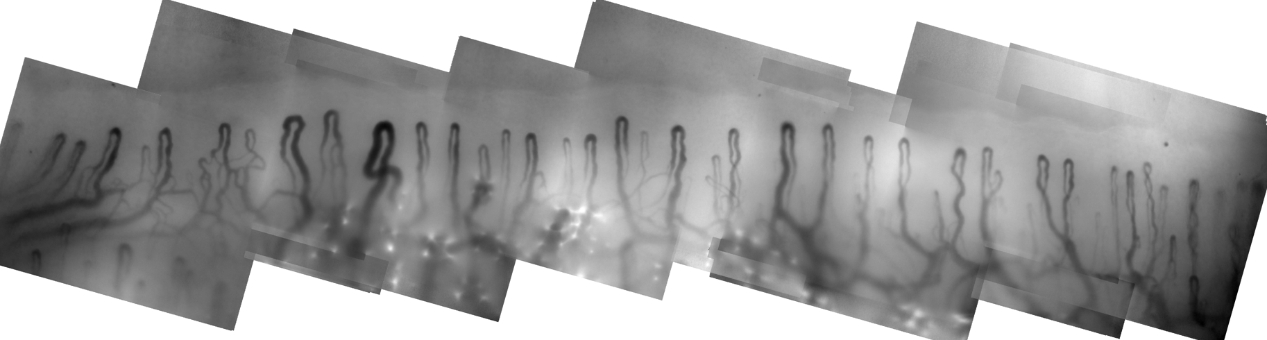
Since July 2011 I have been working on a project funded by the Wellcome Trust developing a new nailfold capillaroscopy system for use in the at Salford Royal NHS Foundation Trust (SRFT). Their existing system uses a video camera attached to a microscope to capture high-magnification images with a spatial resolution of approximately 1 micron per pixel (see figure above). The capillaries in each video frame are only partially captured, as they can only be seen where red blood cells are present. Thus successive video frames are averaged together to provide the best possible image of capillary structure. To enable the full nailfold to be imaged, the microscope and camera are mounted on a high-precision movable 3D stage. By adjusting this stage, several composite frames may be mosaicked together to produce a single panorama (below).
While the system is capable of producing images and has helped the research team lead by Dr Ariane Herrick become an internationally renowned centre of expertise in capillaroscopy (see key publications below), it was developed approximately 15 years ago and naturally parts of the system are no longer state-of-the-art. Our project was tasked with improving the system in three key ways:
- Update the hardware making use of state-of-the-art components
- Write software to automatically analyse nailfold images (both new images and those captured on the old system)
- Use the increase frame rate of the new camera system to measure blood velocity in the capillaries
Update at January 2015:
We are close to completing the project and will shortly be trialling the new system at SRFT.
The content below has temporarily been removed to preserve the anonymity of conference paper submission. I will reinstate it as soon as possible
A new camera system
The video below shows a screen grab of the new system in use during a live recording session. The user controls the 3D motorised platform to which the camera is connected by moving an onscreen joystick
or clicking to where they wish move next in the live-view. Arrow keys or the mouse-wheel move the platform vertically to enable fine-tuned focus. The camera may also be autofocused by pressing the space-bar or double clicking
a location in the live-view. During recording a real-time mosaic of the frames is displayed. When recording stops the full sequence of frames are automatically processed to improve the quality of the mosaic,
selecting only in-focus frames at each spatial location.
The 3D platform can itself be tilted and locked at any angle from its default vertical position to near-horizontal using a motorized actuator, so that the focus plane of the camera can be rotated through approximately 75 degrees.
This enables the imaging of patients with contractures that prevent them from placing their fingers flat against the support platform.
Measuring blood flow
Analysis of blood flow will begin when we have collected sufficient data from the camera trial. Initial results on test sequences are promising.
Key publications
1. Berks, An Automated System for Detecting and Measuring Nailfold Capillaries, MICCAI, LNCS 8673:658-6655, 2014
2. Murray, Non-invasive Imaging Techniques in the Assessment of Scleroderma Spectrum Disorders, Arthritis Care and Research, 61(8), 2009
Some other bits of fun!
Using a technique developed by Dr Phil Tresadern for synthesising capillaries (see publications), I've been creating logos for the 2015
annual meeting of the British Microcirculation Society. A selection of these are shown below...
![]()
![]()
![]()
This website will look much better in a web browser that supports web standards, but it is accessible to any browser or Internet device.