Estimating Skeletal Maturity from X-Rays of the Hand
Steve Adeshina and Tim Cootes
Estimating skeletal maturity plays an important role in the diagnosis of growth and
endocrine disorders. The two main methods examine the
morphology of the bones and the joints of the non-dominant hand in a radiograph.
A significant difference between the bone age and the actual age of a child is an indication of growth abnormalities.
In this project we explored the use of statistical models of shape and appearance for estimating
skeletal maturity.
 |
 |
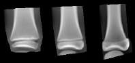 |
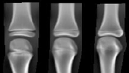
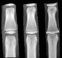
| Metacarpal | Mid.Phalanges | Prox.Phalanges |
Overall the system was able to produce accurate predictions, with average errors of less than one year.
See the publications (and Steve Adeshina's thesis) for details.
Publications
[1] S.Adeshina and T.F.Cootes, "Constructing Part-based Models for Groupwise Registration", Proc. IEEE Int. Symp. on Biomedical Imaging (ISBI) 2010.
[2] S.A.Adeshina, T.F.Cootes and J.E.Adams, "Evaluating the use of Carpal bones for the determination of skeletal maturity for infants", Proc MIUA 2010, pp.279-283
(PDF)
[3] S.A.Adeshina and T.F.Cootes, "Evaluation of Performance of Part-based Models for Groupwise Registration", Proc MIUA 2010, pp.221-225. (Best Poster Prize)
(PDF)
[4] S.A. Adeshina, T.F. Cootes and J.E. Adams
"Evaluating different structures for predicting skeletal maturity using statistical appearance models",
MIUA 2009 (PDF)
[5] S.A. Adeshina, PhD Thesis: "Automatic Determination of Skeletal Maturity using Statistical Models of Appearance", The University of Manchester, 2010. (PDF, 15Mb)