|
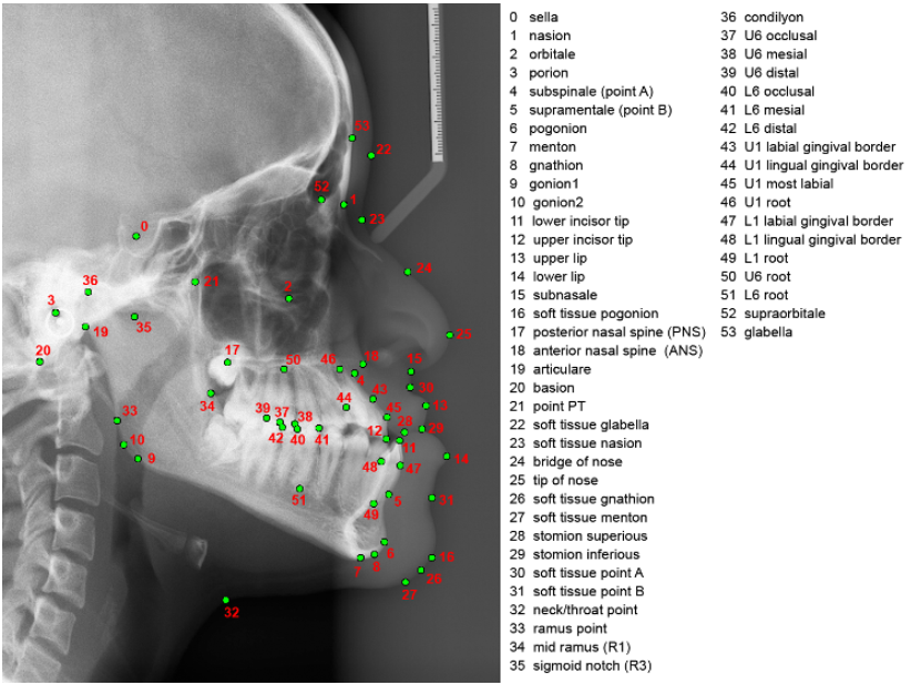
Background: Cephalometric analysis studies the dental and skeletal relationships in the head and is commonly used for orthodontic diagnosis and treatment planning. The identification of cephalometric landmarks on images of the skull allows the quantification and classification of anatomical abnormalities. In clinical practice, the landmarks are placed manually by experienced doctors; a time-consuming and subjective process with inconsistent results. Technology: We have developed a fully automatic landmark annotation system for finding 54 cephalometric landmarks in 2D lateral cephalograms. The system incorporates sophisticated machine learning algorithms to accurately and robustly identify cephalometric landmarks on new unseen data. An earlier version, using a subset of 19 cephalometric landmarks, was awarded the 1st prize at the International Grand Challenge on Automatic Detection of Anatomical Landmarks in Cephalometric X-Rays (Workshop at ISBI 2015). Performance: The 54 points fully automatic system was evaluated on 289 images and achieved a mean point-to-point error of 1.6mm, and for 80% of images the average point-to-point error was within 2.0mm. For the worst performing image the average point-to-point error was 3.5mm. The average runtime per image was less than 1.5 seconds. The performance of our system was shown to be within the inter-observer variability between two clinical experts. Intellectual property: The core technology of the system has been patented (US 9928443, EP 2893491). Opportunity: We are seeking partners to license the software as a plug-in to existing offerings within the dentistry market, including companies that produce and/or sell X-ray machines and companies interested to enter the market. Please contact me if you are interested in trialling the technology and discussing this further. |


|
Publications: Adaptable landmark localisation: applying model transfer learning to a shape model matching system C. Lindner, D. Waring, B. Thiruvenkatachari, K. O'Brien and T. Cootes. C. Lindner, C-W. Wang, C-T. Huang, C-H. Li, S-W. Chang and T. Cootes. A benchmark for comparison of dental radiography analysis algorithms C.-W. Wang, C.-T. Huang, J.-H. Lee, C.-H. Li, S.-W. Chang, M.-J. Siao, T.-M. Lai, B. Ibragimov, T. Vrtovec, O. Ronneberger, P. Fischer, T.F. Cootes and C. Lindner. Fully Automatic Cephalometric Evaluation using Random Forest Regression-Voting C. Lindner and T.F. Cootes.
|