|
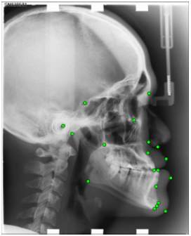
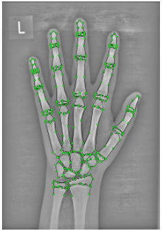
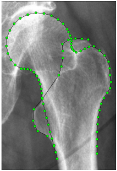
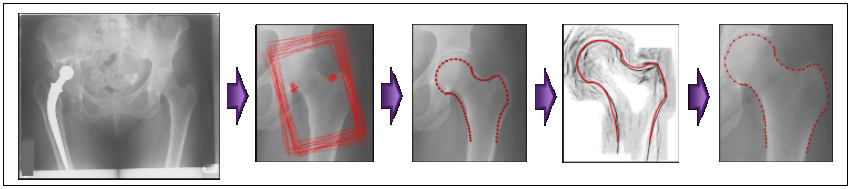
The key goal of this project is to develop robust and accurate systems for locating the outlines of bones and other structures in widely used medical images such as radiographs. A key motivation is to provide a set of tools for the clinical research community to help analyse bone shape and thus better understand, monitor and treat musculoskeletal disease. Such diseases affect about 16% of all adults and more than 30% in the over 65s. The annual total cost of arthritis alone is estimated to be more than £30 billion for the UK. There is increasing clinical interest in studying bone shape, but progress is hampered by the time required to annotate the outlines of structures of interest on the large databases now available. The project will produce both a practical and useful system for clinicians, and new algorithms and insights into tackling fundamental problems in computer vision and medical image analysis.
|
|
Robust Systems for Automated Analysis of Structures in 2D Medical |


|
Claudia Lindner, Richard Hodgson, Judy Adams and Tim Cootes |
|
Grand Challenge on Automatic Detection of Anatomical Landmarks in Cephalometric X-Rays (Workshop at ISBI2015): First prize Fully Automatic Cephalometric Evaluation using Random Forest Regression-Voting C. Lindner and T.F. Cootes. Proceedings of the IEEE International Symposium on Biomedical Imaging (ISBI) 2015 – Grand Challenges in Dental X-ray Image Analysis – Automated Detection and Analysis for Diagnosis in Cephalometric X-ray Image, 2015. |